Raman Microscopy
Raman Microscopy
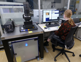
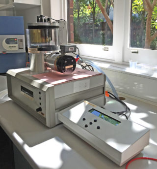
A WITec alpha300R Confocal Raman Microscope for two excitation lasers with wavelengths of 532 and 785 nm was installed in the LGIM Facility in May 2024. Raman microscopy uses inelastic photon scattering to identify characteristic molecular vibrations in matter. Excitation of electrons by intense laser beams creates bond-specific emissions that can be spectrally resolved in the scattered light. Laser beam interaction with the target material is non-destructive and spatially selective at micrometre lateral resolution, depending on excitation wavelength and numerical aperture of the microscope objective. Three-dimensional mapping is possible in transparent materials at sub-μm depth resolution in confocal illumination. Available objectives are 10× (numerical aperture NA 0.95, working distance WD 9.3 mm), 40× (NA 0.6; WD 3.6 mm), 50× (bright-/darkfield NA 0.8; WD 0.6 mm), and 100× (NA 0.9; WD 1.0 mm). Raman microscopy requires minimal sample preparation, with no need for coating, polishing, or vacuum-stable mounting.
Raman Current Applications
The Raman microscopy capability enhances the isotopic and compositional analytical capability of LG-SIMS by providing fine-scale structural analysis of materials. Rapid, non-destructive, and spatially selective Raman analyses are applied to research questions in the Earth and planetary sciences, and other disciplines concerned with material structural properties.