Microscopy & Microanalysis Facility
The MMF houses a broad range of advanced microanalysis instrumentation providing high quality chemical, mineralogical and microstructural information, and high resolution images for research and technical publications. The facility staff have expertise in Materials and Earth Science research which is used to support both academic and applied research in science, engineering and biological disciplines across a broad range of academic projects and in Western Australian industries, particularly in minerals and energy sectors.
Techniques/Capabilities
TIMA (Tescan Integrated Mineral Analyzer)
- Automatic PGM (platinum-group minerals) search on multiple samples at the same time (22 x 25-30mm mounts or 9 x thin sections or polished sections)
- Modal abundance
- Size-by-size liberation
- Mineral association
- Colour CL imaging
SEM
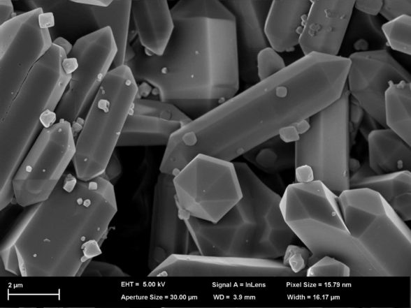
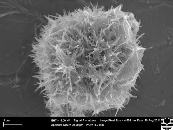
- High resolution imaging with secondary electrons (SE), backscattered electrons (BSE) and cathodoluminescence (CL)
- Panorama imaging in SE, BSE and CL modes
- Qualitative and quantitative energy-dispersive x-ray spectroscopy (EDS) elemental analysis
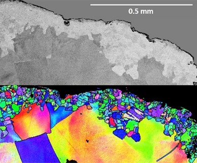
- Electron backscattered diffraction (EBSD) for phase and orientation analysis
- Large area mapping (LAM) for EDS and EBSD
- Transmission Kikuchi Diffraction (TKD)
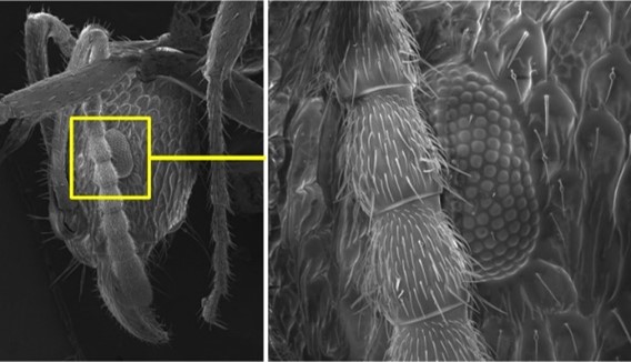
- Variable pressure (VP) imaging of delicate/uncoated/moist samples
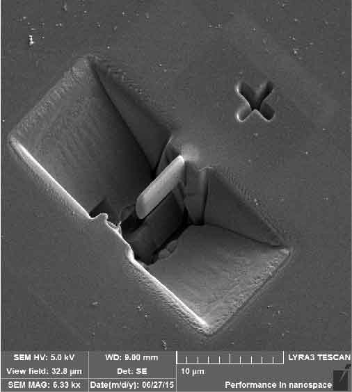
- Ga+ Focused Ion Beam (FIB) sample manipulation for sectioning, in-situ lift-out and 3D imaging (via FIB Facility)
- Light microscopy, including low magnification stereomicroscopy
- Sample preparation equipment including carbon and platinum coaters
- Mounting/polishing equipment
TEM
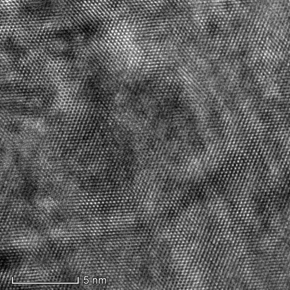
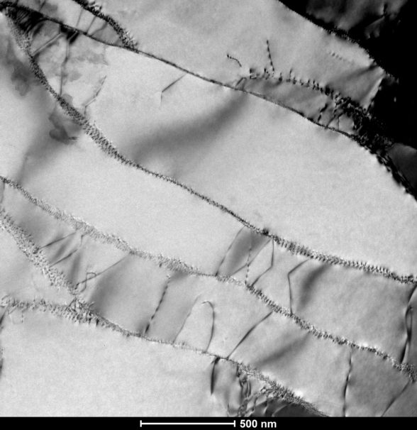
- Atomic lattice imaging
- STEM imaging in HAADF, BF and DF modes
- Qualitative and quantitative energy-dispersive x-ray spectroscopy (EDS) elemental analysis
- Single tilt and low background double tilt holders
- TEM, STEM and EDS tomography
- Electron diffraction by SAD and CBED
INSTRUMENTS
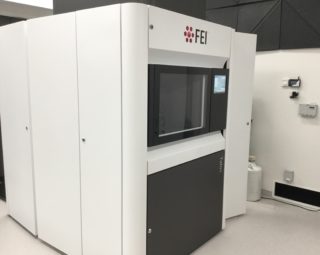
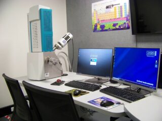
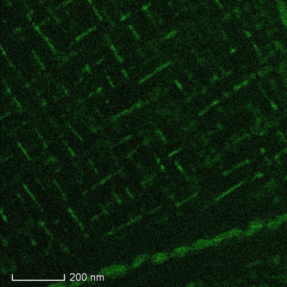
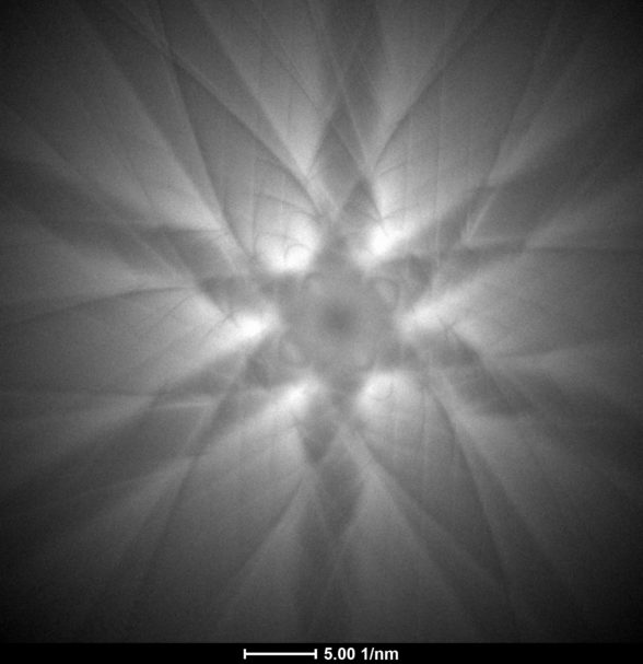
TALOS FEG TEM
The FEI Talos F200X G2 field emission gun (FEG) transmission electron microscope (TEM) is built on an advanced operating platform for atomic lattice resolution imaging and nanoscale microanalysis. The TEM is equipped with highly sensitive Super-X EDS detectors for elemental analysis. This instrument is capable of 3D tomography investigations in all modes of operation (TEM, STEM & EDS).
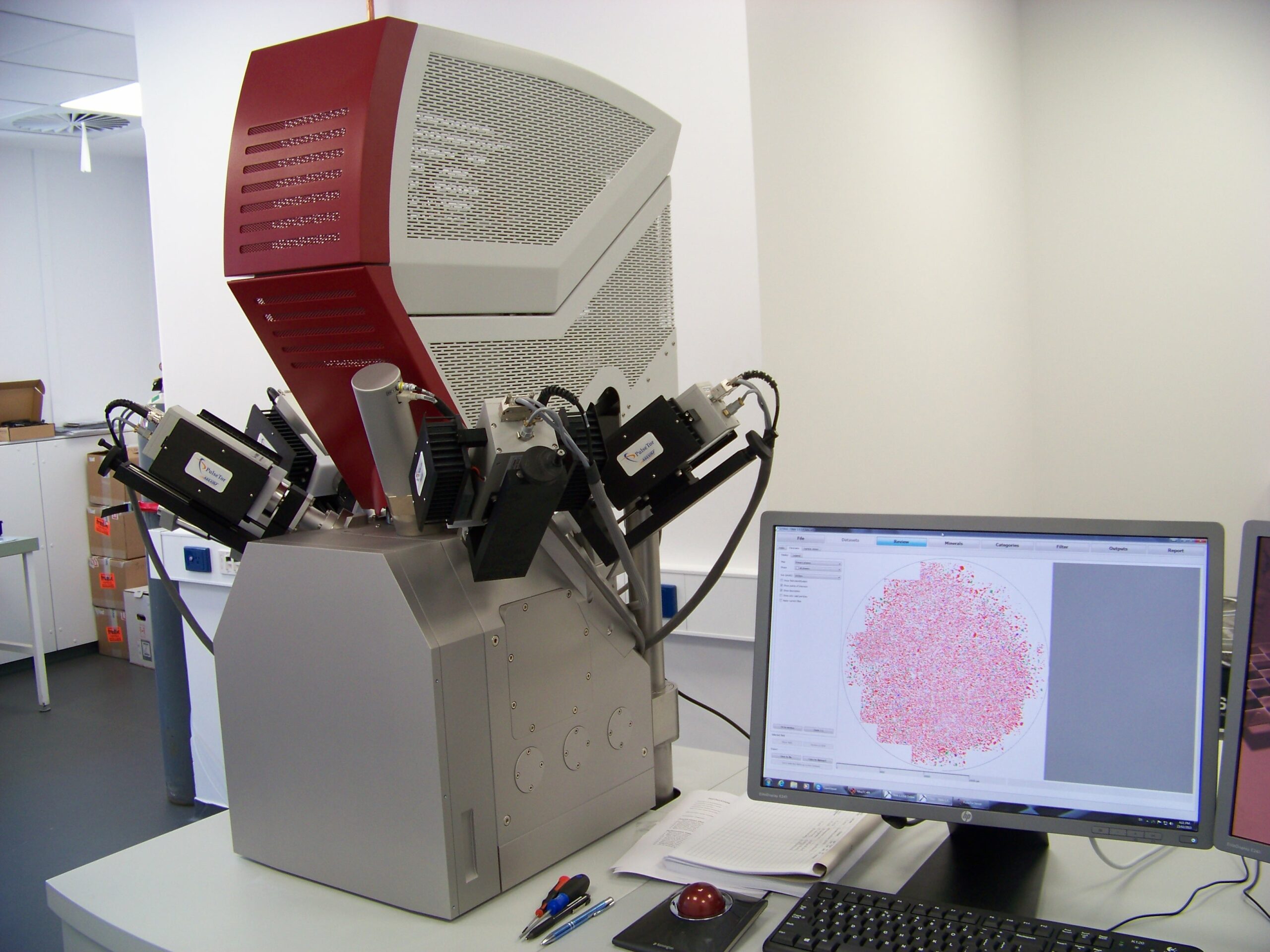
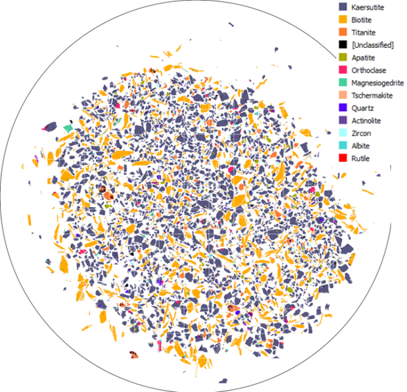
TIMA (Automated Mineralogy)
The Tescan Integrated Mineral Analyzer (TIMA GM) is a fully automated, high throughput, analytical Field Emission Scanning Electron Microscope (FESEM) used to obtain sample composition and morphology. The TIMA measures modal abundance, size-by-size liberation and mineral association, and performs a PGM search automatically on multiple samples of grain mounts and thin or polished sections. It is equipped with an ultra-fast YAG scintillator BSE detector, complemented with four silicon drift EDS X-ray detectors that cover a maximum solid angle of X-ray data acquisition for high throughput analysis. Collection of EDS data is synchronised with a scanning system and BSE signal acquisition using integrated hardware.
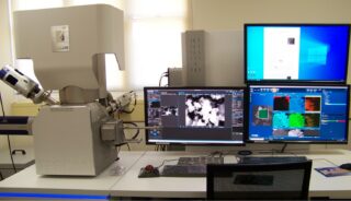
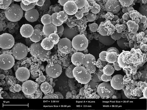
CLARA FESEM
The Tescan Clara is a field emission scanning electron microscope (FESEM) with a comprehensive range of detectors to enable research in the fields of earth, life, forensic and materials sciences. Its special features are its higher beam current (compatible with Oxford Symmetry EBSD speed) and its high sensitivity EDS detector that enables very low kV and high spatial resolution microanalysis, coupled with low kV imaging that is also suitable for non-conductive samples.
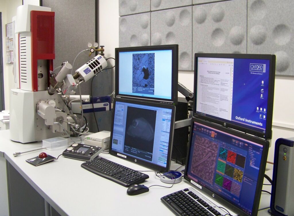
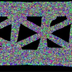
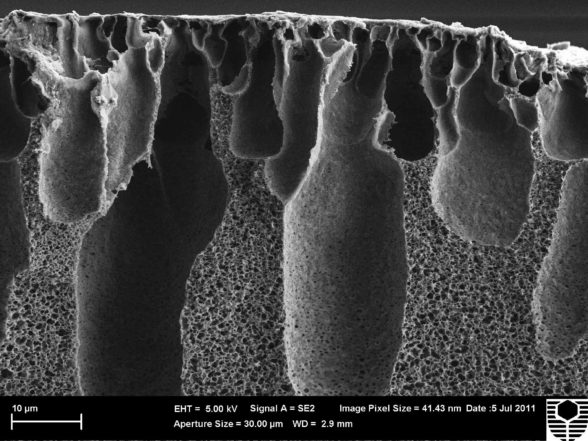
MIRA VP-FESEM
The Tescan Mira3 is a variable pressure field emission scanning electron microscope (VP-FESEM) that features sensitive EDS and EBSD detectors and integrated software for high quality microstructural analysis of crystalline samples. It has a full complement of detectors (SE, BSE, In-beam SE/BSE and CL). Variable pressure enables analysis of porous or moist samples without need for coating.
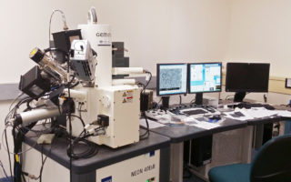
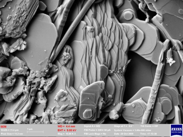
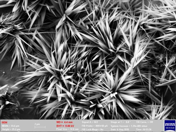
NEON DUAL-BEAM FESEM
The Zeiss Neon 40EsB is a dual-beam field emission scanning electron microscope (FESEM). This instrument combines extremely high resolution imaging with regular EDS and EBSD.
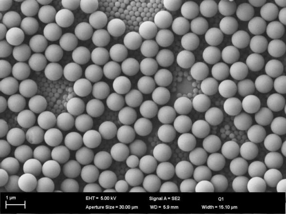
VEGA VP-SEM
The Tescan Vega3 is a variable pressure scanning electron microscope (VP-SEM) with a tungsten filament. It is suitable for lower magnification microstructural analysis at high vacuum, or for the analysis of non-conductive/hydrated samples at lower vacuum, with a smallest particle size of approximately one micrometre.
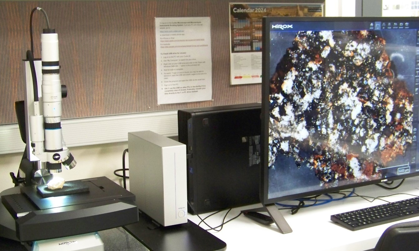
HIROX DIGITAL LIGHT MICROSCOPE
The Hirox (HRX-01) is a digital light microscope that can acquire 3D and 2D images using multiple focus points. Using its fully motorised lenses and stage, the Hirox is capable of high resolution, true colour imaging (with a scale bar of 10 µm) while also being able to cover large areas (up to 100 cm2) at lower magnifications by stitching images together. The software also facilitates multiple post-processing functions including roughness, depth and area measurements, as well as profilometry.
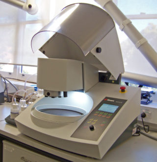
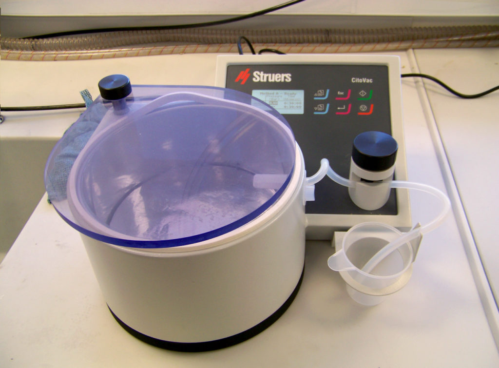
Sample Preparation Laboratories
The MMF has a suite of equipment that includes a broad beam ion mill, coaters and desiccator systems that are used to prepare samples for electron microscopy. It also has equipment that enables vacuum mount impregnation and both manual and automated polishing, which is often required to prepare samples for electron microscopy and is essential for TIMA and quantitative EDS analyses.
Applications
Collaborative research, training and education, and consultancy in the fields of:
Earth Science
- Quantification of sample microstructure and mineralogy
- Mineral liberation analysis, process optimisation, remediation, and searches for precious metals and rare earths specifically addressed by TIMA
- High resolution EDS and EBSD mapping of geological samples
- Characterisation of crystallographic preferred orientations in deformed minerals
- Formation and deformation of ore minerals and ore bodies
Materials Science
- Investigation of corrosion products, asbestos fibres and building materials
- Characterisation of crystallographic texture and orientation
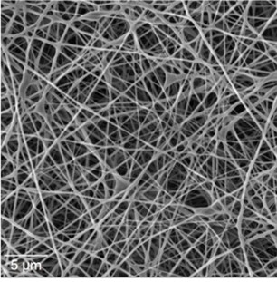
- Analysis of morphology, particle size and porosity
- Fracture and damage analysis
Chemical Science
- High resolution imaging and chemical analysis of electrodes, nano-particles and reaction products
- Crystallographic analysis of complex synthetic samples
Life Science
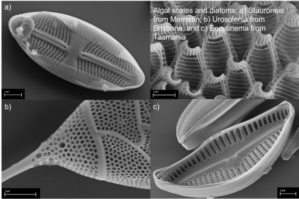
- Microstructural analysis of biological samples, including those that are not suitable for either high vacuum or high voltage due to their hydration and/or fragility

Access to MMF
Registration
- To register or enquire about rates please contact MMF via email.
Access to Instrumentation
- Curtin staff/students and associates – There is a Registration Fee for all new users, which also covers a new user meeting to discuss your access requirements and appropriate sample preparation. It is compulsory to attend the MMF Induction if you will be using the Preparation Laboratory or any of the instruments unsupervised. After completing training, regular users can book MMF instruments via our online booking system; irregular users will need to email us to arrange a suitable time when they, a technician and the instrument are available.
- All other users – Please email us to discuss your access.